Home Sleep Apnea Testing vs In-Lab Polysomnography: Accuracy Insights
Home sleep apnea testing (HSAT) and in-lab polysomnography (PSG) serve as pivotal diagnostic tools in sleep medicine, yet their accuracy diverges significantly. While HSAT offers convenience and cost-effectiveness, in-lab PSG remains the gold standard due to its comprehensive assessment of sleep architecture and neurophysiological parameters. Understanding these differences is crucial for optimizing patient outcomes in sleep-related disorders.
Neurochemical & Stage Physiology of Sleep Apnea
The neurochemical landscape during sleep is intricately linked to the stages of sleep, particularly in the context of sleep apnea. During rapid eye movement (REM) sleep, the brain exhibits heightened activity, characterized by the release of neurotransmitters such as acetylcholine, which facilitate vivid dreaming and cognitive restoration. Conversely, non-REM (NREM) sleep, particularly stages 3 and 4, is dominated by slow-wave activity (SWA) and the release of growth hormone, essential for physical recovery and memory consolidation. Disruptions in these stages due to apnea events can lead to fragmented sleep, affecting cognitive functions and emotional regulation.
In patients with obstructive sleep apnea (OSA), the repeated episodes of airway obstruction lead to intermittent hypoxia, triggering a cascade of neurochemical responses. This includes the activation of the sympathetic nervous system, resulting in elevated levels of norepinephrine and cortisol, which can impair sleep quality and cognitive performance. Studies have shown that individuals with untreated OSA exhibit reduced slow-wave sleep percentages, often dropping below the critical threshold of 20% of total sleep time, which is vital for effective glymphatic beta-amyloid clearance and overall brain health Glymphatic Beta-Amyloid Clearance Rates: Deep Sleep Insights.
Clinical Diagnostic Endpoints: AHI, Latency, and Slow-Wave Percentage
Accurate diagnosis of sleep apnea hinges on several clinical endpoints, including the Apnea-Hypopnea Index (AHI), sleep latency, and the percentage of slow-wave sleep. The AHI quantifies the severity of sleep apnea by measuring the number of apneas and hypopneas per hour of sleep. An AHI of 5-15 indicates mild OSA, 15-30 denotes moderate OSA, and over 30 signifies severe OSA. In contrast, HSAT typically captures a limited range of data, often yielding AHI values that may underestimate the severity of the condition due to its inability to monitor sleep stages comprehensively.
Sleep latency, defined as the time taken to transition from full wakefulness to sleep, is another critical metric. Normal sleep latency ranges from 10 to 20 minutes; however, prolonged latency may indicate underlying sleep disorders or excessive daytime sleepiness. In patients with OSA, sleep latency can be significantly affected, with studies indicating an average latency increase of 30% in those with untreated conditions. Furthermore, the percentage of slow-wave sleep is crucial for cognitive restoration, with healthy adults typically achieving 20-25% of total sleep time in this stage. In OSA patients, this percentage often drops to below 15%, further exacerbating cognitive deficits.
Intervention Comparison: HSAT vs. PSG
| Parameter | Home Sleep Apnea Testing (HSAT) | In-Lab Polysomnography (PSG) |
|---|---|---|
| Diagnostic Accuracy | Moderate (sensitivity ~70-80%) | High (sensitivity ~90-95%) |
| Cost | Lower ($150-$300) | Higher ($1,000-$3,000) |
| Data Collected | Limited (heart rate, airflow, oxygen saturation) | Comprehensive (EEG, EOG, EMG, respiratory patterns) |
| Patient Comfort | High (at-home setting) | Variable (hospital setting) |
| Time to Results | Short (1-2 weeks) | Longer (2-4 weeks) |
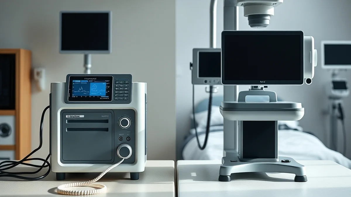
When comparing HSAT and PSG, the differences in diagnostic accuracy and data collection are striking. HSAT primarily measures basic physiological parameters, leading to a sensitivity range of approximately 70-80%. In contrast, PSG’s comprehensive approach, which includes electroencephalography (EEG) and electromyography (EMG), enhances its sensitivity to 90-95%. This disparity is critical, as accurate diagnosis directly influences treatment efficacy and patient outcomes.
Moreover, while HSAT is more cost-effective and allows for greater patient comfort, it may overlook complex sleep disorders that require detailed analysis. The choice between these two diagnostic modalities should be guided by clinical indications, patient history, and the need for comprehensive sleep architecture assessment.
Pharmacological and Sedative Confounding Traps
Pharmacological agents, including sedatives and antidepressants, can significantly confound sleep study results. Medications such as benzodiazepines may enhance sleep continuity but can suppress REM sleep and slow-wave sleep, leading to an inaccurate assessment of sleep apnea severity. Understanding the impact of these drugs is essential for interpreting diagnostic outcomes.
In the realm of sleep medicine, the influence of pharmacological agents on sleep architecture cannot be overstated. Sedatives, while often prescribed to manage insomnia, can mask the underlying symptoms of sleep apnea. For instance, benzodiazepines may increase total sleep time but can reduce the proportion of REM and deep sleep, leading to a false sense of security regarding sleep quality. This suppression of critical sleep stages can result in an inaccurate AHI, potentially delaying appropriate intervention.
Furthermore, antidepressants such as selective serotonin reuptake inhibitors (SSRIs) can alter sleep patterns, often leading to increased awakenings and reduced sleep efficiency. These effects complicate the interpretation of HSAT and PSG results, necessitating careful consideration of a patient’s medication regimen prior to testing. Clinicians must balance the need for pharmacological intervention with the potential for confounding effects on sleep diagnostics.
Related Aegis Lab Briefings: Explore deeper benchmarks in Positional Obstructive Sleep Apnea vs Mandibular Advancement Devices.
Evening Hygiene & Clinical Escalation Steps
Establishing a robust evening hygiene routine is essential for optimizing sleep quality and mitigating the effects of sleep apnea. Patients should be encouraged to maintain a consistent sleep schedule, aiming for 7-9 hours of sleep per night, and to create a conducive sleep environment free from distractions. Limiting exposure to screens and blue light in the hour leading up to bedtime can enhance melatonin production, promoting the onset of sleep. Additionally, avoiding stimulants such as caffeine and nicotine in the evening is crucial for reducing sleep latency and improving overall sleep quality.
In cases where home sleep testing indicates moderate to severe sleep apnea, clinical escalation steps should be implemented. This may include referral for in-lab PSG for comprehensive evaluation, consideration of continuous positive airway pressure (CPAP) therapy, or lifestyle interventions such as weight management and positional therapy. Regular follow-ups are essential to monitor treatment efficacy and adjust interventions as needed, ensuring that patients receive the most effective care tailored to their specific needs.
Protecting deep slow-wave sleep consistency and airway patency preserves long-term cognitive reserve.